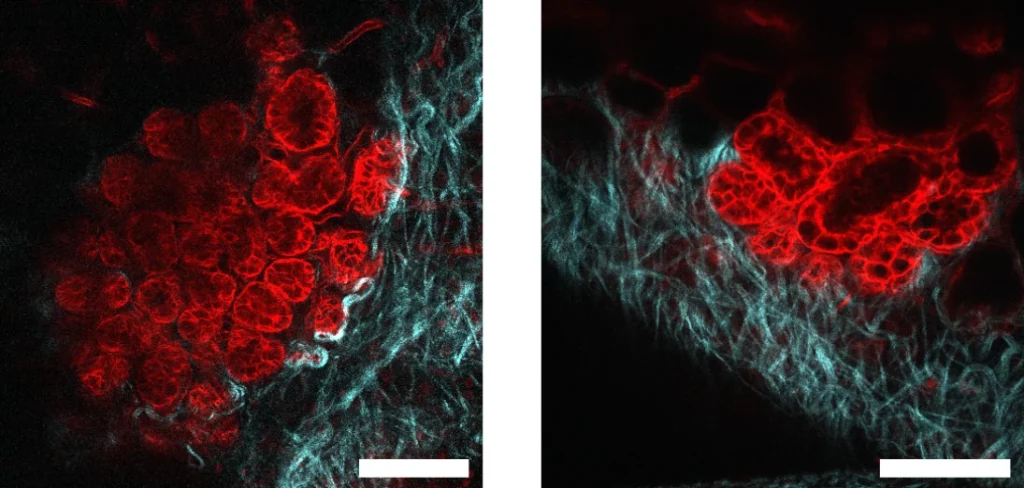
Credit: Emma Anisman, Igor Luzhansky, Washington University in Saint Louis In Vivo Imaging Core.
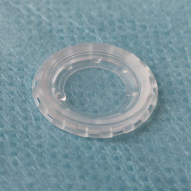
Intravital imaging makes it possible to follow the mechanisms of life over time on the same individuals, thus reducing the animal use while powering the data. That is why we developed our PDMS intravital window!
Did you know that as like humans, every animal is different? Of course… which is why, when experiments cannot be conduct in dish, in vivo experiments require to cull batches of animals to obtain conclusive results…
Safe, Flexible and Water Clear
Intravital imaging remains a heavy procedure for the animal. By developing our PDMS window, we aimed to :
- Reduce the weight up to 10x compare to conventional titanium/glass windows.
- Reduce discomfort and pain by providing soft and flexible device.
- Reduce the risk due to the reuse by providing disposable device
- Improve experiment by allowing optimal 2PM imaging (water close refractive index) and in-situ operations thanks to its injection port.


Perform the Quickest Surgery with our Intravital Window
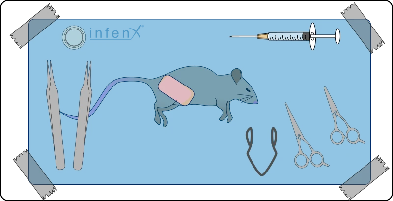
The failure of a surgical procedure is most often due to its duration. For the researcher, for the animal, for the results, we sought to reduce the duration of the operation as much as possible. No sutures… high tolerance… obtain subcutaneous access in less than 5 minutes from the first incision to sealing the window…
Imagine what was not possible before
Thanks to its flexibility, you can now implant a window where it was not possible before. Neck, leg, belly, our flexible window made in PDMS is optimal for flexible region… Isn’t it obvious?


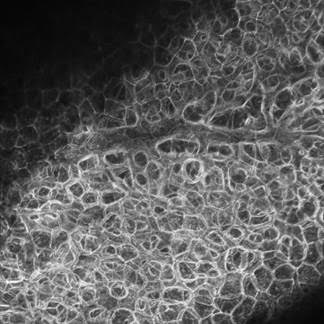
Liver – mTomato
References
Guillaume Jacquemin et al. ,Longitudinal high-resolution imaging through a flexible intravital imaging window.Sci. Adv.7,eabg7663(2021). DOI:10.1126/sciadv.abg7663
We aims to support any young scientist to reach there goal and money should never be the limit – feel free to contact us to share your projects and discuss how we can help you